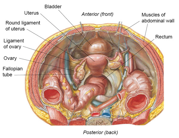

Pouch of Douglas: The anatomic space between the rectum and uterus. It can be palpated via a digital rectal exam. It is an important anatomic area because it can be a collecting site of blood (from ruptured ectopic pregnancy), pus (from pelvic inflammatory disease), malignant cells (from ovarian cancer), or endometrial implants (from endometriosis).
Course of the female ureters: Cross over the bifurcation of the common iliac arteries, then lateral to the internal iliac arteries. They enter the retroperitoneal space in the uterosacral ligaments, then the cardinal ligaments, and dive under the uterine artery.
The ureter is sometimes injured in surgeries involving ligation of the uterine artery because of their anatomic proximity. Surgeons must be wary!
Mnemonic: Water under the bridge.

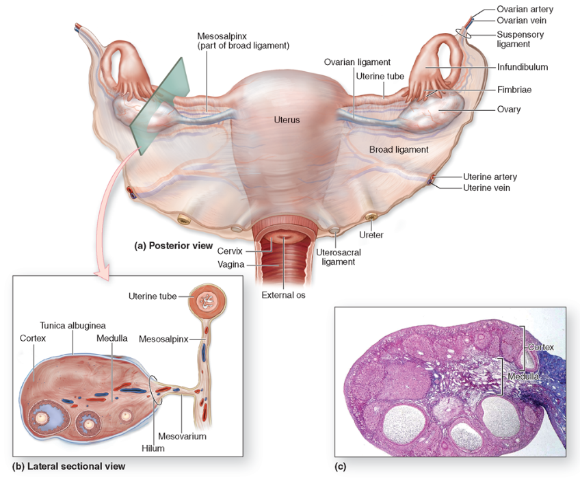
Coronal section of pelvis demonstrating the ligaments of the female reproductive tract.
Infundibulopelvic ligament: Also known as the suspensory ligament of ovaries. Connects the ovaries to the pelvic wall and contains the ovarian blood vessels.
Ovarian ligament: Connects the ovaries to the lateral surface of the uterus. It does not contain any vessels.
Round ligament: Connects the uterine fundus to the labia majora by passing through the deep inguinal ring. The round ligament is a derivative of the embryologic gubernaculum.
Cardinal ligament: Connects the cervix to the pelvic side wall. It contains the uterine blood vessels.
Broad ligament: Connects the ovaries, fallopian tubes, and uterus to the pelvic floor and side wall.
Clinical Pearls: DON’T cut the: I nfundibulopelvic ligament, C ardinal ligament, or U reter OR your patient will end up in the ICU !
The pouch of Douglas can be a collecting site for blood, pus, malignant cells, or endometrial implants.
The menstrual cycle occurs throughout the reproductive period, with an average cycle length of approximately 28 days. These cycles cease at about the fifth decade of life (menopause), at which time the reproductive organs become atrophic.
Attachments of the Ovary:
- infundibulopelvic ligament - fallopian tube attaches to the ovary is connected to it by.
- The other extremity is called the uterine extremity. It points downward, and it is attached to the uterus via the ovarian ligament.
Female Gonadal Venous and Lymphatic Drainage
The venous drainage of the right ovary or testis is into the right ovarian and then into the inferior vena cava. The venous drainage of the left ovary is into the left ovarian or testicular vein, then into the left renal vein, and then into the inferior vena cava. The extra step in left testicular venous drainage causes increased hydrostatic pressure. That is why varicoceles are more common on the left side.

Vascular and lymphatic system of the reproductive tract.
++++++++++++++++++++++++++++++++++++++++++++
The lymphatic drainage from the ovaries is into the paraaortic nodes (this drainage pattern occurs because the ovaries and testes descended from the abdomen with their blood source from the aorta). The lymphatic drainage of the distal third of the vagina is into the superficial inguinal nodes. The uterus and proximal two thirds of the vagina drain into the external iliac, obturator, and hypogastric nodes.
Reference